Radiation can damage some types of normal tissue more easily than others. For example, the reproductive organs (testicles and ovaries) are more sensitive to radiation than bones. The radiation oncologist takes all of this information into account during treatment planning.
If an area of the body has previously been treated with radiation therapy, a patient may not be able to have radiation therapy to that area a second time, depending on how much radiation was given during the initial treatment. If one area of the body has already received the maximum safe lifetime dose of radiation, another area might still be treated with radiation therapy if the distance between the two areas is large enough.
The area selected for treatment usually includes the whole tumor plus a small amount of normal tissue surrounding the tumor. The normal tissue is treated for two main reasons:
• To take into account body movement from breathing and normal movement of the organs within the body, which can change the location of a tumor between treatments.
• To reduce the likelihood of tumor recurrence from cancer cells that have spread to the normal tissue next to the tumor (called microscopic local spread).
How is radiation therapy given to patients?
Radiation can come from a machine outside the body (external-beam radiation therapy) or from radioactive material placed in the body near cancer cells (internal radiation therapy, more commonly called brachytherapy). Systemic radiation therapy uses a radioactive substance, given by mouth or into a vein, that travels in the blood to tissues throughout the body.
The type of radiation therapy prescribed by a radiation oncologist depends on many factors, including:
- The type of cancer.
- The size of the cancer.
- The cancer’s location in the body.
- How close the cancer is to normal tissues that are sensitive to radiation.
- How far into the body the radiation needs to travel.
- The patient’s general health and medical history.
- Whether the patient will have other types of cancer treatment.
- Other factors, such as the patient’s age and other medical conditions.
External-beam radiation therapy
External-beam radiation therapy is most often delivered in the form of photon beams (either x-rays or gamma rays) (1). A photon is the basic unit of light and other forms of electromagnetic radiation. It can be thought of as a bundle of energy. The amount of energy in a photon can vary. For example, the photons in gamma rays have the highest energy, followed by the photons in x-rays.
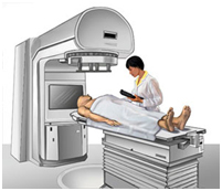
 Linear Accelerator Used for External-beam Radiation Therapy. Many types of external-beam radiation therapy are delivered using a machine called a linear accelerator (also called a LINAC). A LINAC uses electricity to form a stream of fast-moving subatomic particles. This creates high-energy radiation that may be used to treat cancer.
Many types of external-beam radiation therapy are delivered using a machine called a linear accelerator (also called a LINAC). A LINAC uses electricity to form a stream of fast-moving subatomic particles. This creates high-energy radiation that may be used to treat cancer.
Patients usually receive external-beam radiation therapy in daily treatment sessions over the course of several weeks (see Question 7). The number of treatment sessions depends on many factors, including the total radiation dose that will be given.
One of the most common types of external-beam radiation therapy is called 3-dimensional conformal radiation therapy (3D-CRT). 3D-CRT uses very sophisticated computer software and advanced treatment machines to deliver radiation to very precisely shaped target areas.
Many other methods of external-beam radiation therapy are currently being tested and used in cancer treatment. These methods include:
Intensity-modulated radiation therapy (IMRT): IMRT uses hundreds of tiny radiation beam-shaping devices, called collimators, to deliver a single dose of radiation (2). The collimators can be stationary or can move during treatment, allowing the intensity of the radiation beams to change during treatment sessions. This kind of dose modulation allows different areas of a tumor or nearby tissues to receive different doses of radiation.
Unlike other types of radiation therapy, IMRT is planned in reverse (called inverse treatment planning). In inverse treatment planning, the radiation oncologist chooses the radiation doses to different areas of the tumor and surrounding tissue, and then a high-powered computer program calculates the required number of beams and angles of the radiation treatment (3). In contrast, during traditional (forward) treatment planning, the radiation oncologist chooses the number and angles of the radiation beams in advance and computers calculate how much dose will be delivered from each of the planned beams.
The goal of IMRT is to increase the radiation dose to the areas that need it and reduce radiation exposure to specific sensitive areas of surrounding normal tissue. Compared with 3D-CRT, IMRT can reduce the risk of some side effects, such as damage to the salivary glands (which can cause dry mouth, or xerostomia), when the head and neck are treated with radiation therapy (4). However, with IMRT, a larger volume of normal tissue overall is exposed to radiation. Whether IMRT leads to improved control of tumor growth and better survival compared with 3D-CRT is not yet known (4).
Image-guided radiation therapy (IGRT): In IGRT, repeated imaging scans (CT, MRI, or PET) are performed during treatment. These imaging scans are processed by computers to identify changes in a tumor’s size and location due to treatment and to allow the position of the patient or the planned radiation dose to be adjusted during treatment as needed. Repeated imaging can increase the accuracy of radiation treatment and may allow reductions in the planned volume of tissue to be treated, thereby decreasing the total radiation dose to normal tissue (5).