From coffee shop to fine dining: Second-gen owners of The Curry Club Signature on frog leg biryani & reinventing Singapore Indian cuisine
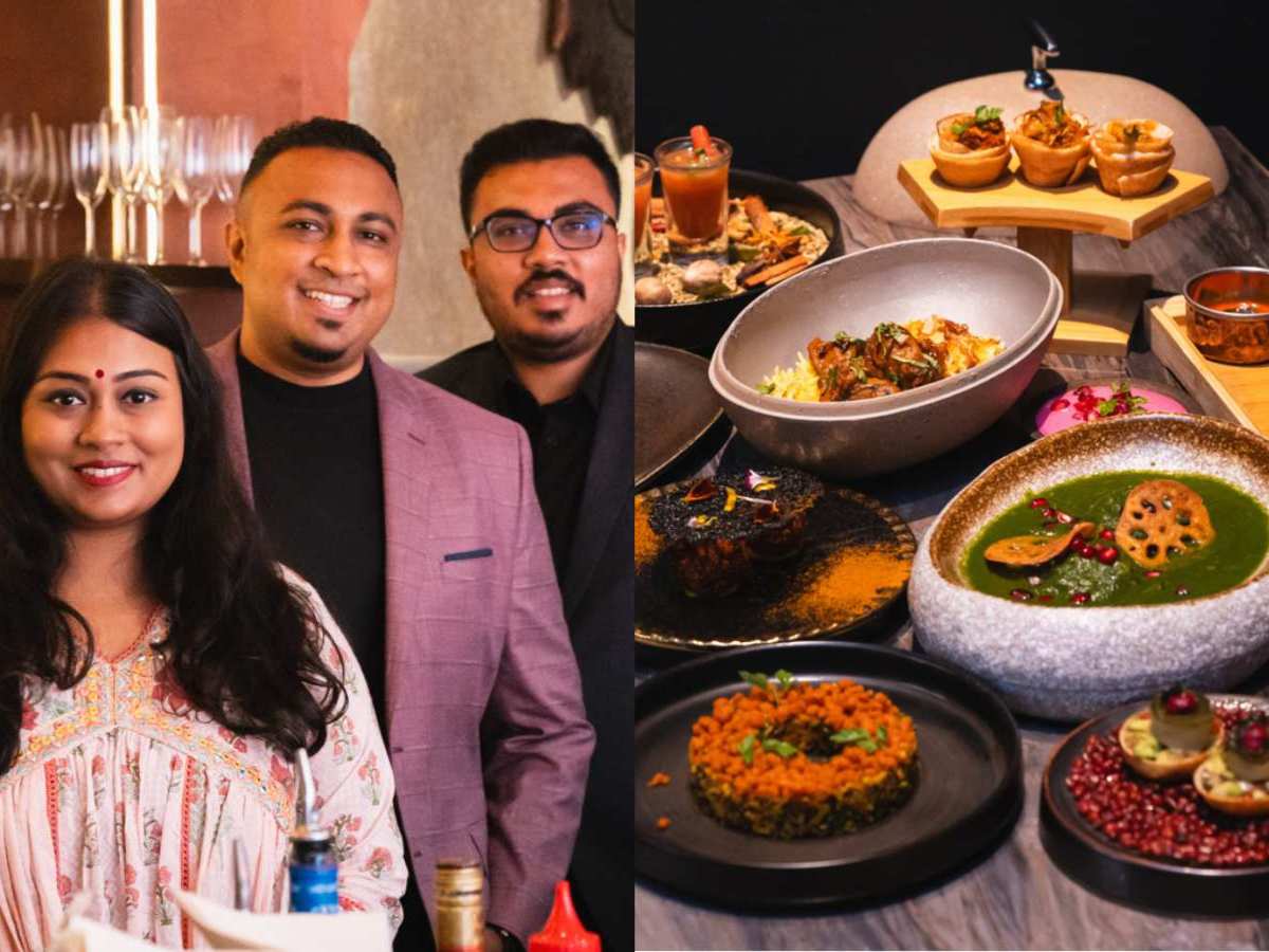
- The Curry Club Signature at Fort Canning is a well-loved family business that’s run by four siblings
- Raakesh Raj, 36, is the founder of the restaurant, where he oversees the restaurant menu and brand
- Come here for one-of-a-kind, contemporary South Indian fare, chiefly its signature frog leg biryani
Nestled along the quaint Mohamed Sultan Road lies The Curry Club Signature, a modish outfit with a stellar reputation for contemporary South Indian fare.
It’s a proud and successful venture between four close-knit siblings: Raakesh, Shaleni, Vigneraj and Tushant Rajendran (Raj).
Raakesh, 36, is the founder of The Curry Club Signature, where he engineers the restaurant menu and brand concept. His siblings are responsible for daily operations, business accounts and marketing efforts.
Together, they ensure that service runs smoothly, the food tastes delicious and all diners leave with a satisfied tummy.
The Curry Club Signature is the newly rebranded concept of gastrobar The Curry Club, which was founded during the peak of the Covid pandemic in 2020.
And since its brand overhaul in September this year, the fine dining establishment has quickly become a hotspot for modern Indian dishes that are well-loved for their rich, saporous and bold flavours.
The siblings take pride in delivering scrumptious food and nothing less. Each dish is to be executed perfectly and its flavours must be consistent.
“It’s not about the money,” Raakesh says. “But rather, we ensure that the best quality is given to the customers.” He adds that this was their father’s motto when working in F&B.
Rajendran Rajoo, 66, continues to be an inspirational figure in their lives — he taught his children the ins and outs of working in a kitchen.
Since then, they’ve all followed his footsteps into the F&B industry.
Now, the four siblings aspire to elevate Singaporean Indian cuisine and bring the family business to greater heights — one dish at a time.

How the family business started
But how did the Raj siblings get into the F&B trade?
It all began when a friend presented Raj Senior with an opportunity to take over the now-defunct Maraj Restaurant at Verdun House in 2006.
Says Raakesh: “He was getting old and couldn’t run the shop anymore, so he approached my dad to take over the business.”
Raj Senior had always wanted to run a business of his own, so it only seemed natural to accept.
Specialising in Indian street delicacies, Maraj was a casual “mamak-style” eatery that operated from 2006 to 2022.
During its sixteen-year tenure, the siblings assumed different roles. Raakesh was tasked with dishing out mee goreng and roti prata, while his brothers manned the cashier. It was where the siblings learnt the ropes of working at a fast-paced cafe, as well as vital cooking skills.
Maraj enjoyed brisk business. It was invited to be the in-house caterer for the Singapore Indian Association in 2008 and proceeded to hold the position for the next 11 years.
“People know us for our food and amazing flavour profiles,” says Raakesh, smiling.
How The Curry Club Signature came to be

Over the years, Maraj became a hit with the masses. The family began to expand their front-of-house team and freed up the family to pursue their own interests.
“The business was running on autopilot by then,” recalls Raakesh.
For instance, Shaleni and Vigneraj went into banking and Tushant enlisted in the army, while Raakesh delved into sales. But the latter soon discovered a newfound passion for the culinary arts and enrolled in a WSQ diploma course at At-Sunrice GlobalChef Academy in 2015. He was 28 then.
Two years later, he set off for New Zealand to pursue a bachelor’s degree in hospitality management. Upon graduating, he clinched a one-year work visa at the prestigious The Langham Hotel in London — one of the top hotels in the world.
“Working at The Langham for two years allowed me to see things in a different light,” Raakesh says. “You learn how to master a certain service etiquette.”
But becoming a chef was not in the cards — he wanted to be an entrepreneur instead.
Being able to refine menus, innovate new brand concepts and network with others was what Raakesh was truly interested in. He preferred to be a part of the action, instead of operating behind the scenes in the kitchen.
An offer to join The Langham Hotel full-time came in 2018, but he found himself turning it down.
Says Raakesh: “Now that I had good exposure in the trade, my dad wanted me to come back so they could start a new family business.”
And that was how The Curry Club was born, a no-frills bistro with a focus on traditional South Indian fare that catered to the local Indian community. The dishes were prepared with generational homemade recipes, courtesy of Raakesh’s grandmother.
After three years, the restaurant rebranded to The Curry Club Signature, with Raakesh at its helm. “We saw a niche in creating a casual fine dining establishment — something not too casual, yet not too serious,” he adds.
They wanted to build a place with elevated Indian food and a great selection of wines, cocktails and dishes that could appeal to diners beyond the Indian community.
The whole family now works full-time at the restaurant, but working alongside his loved ones hasn’t been all sunshine and rainbows for Raakesh.
They don’t always agree.
“But we make decisions together,” he says. “We take votes and move forward.”
There have also been moments when the siblings wanted to throw in the towel.
One instance was when renovation works were delayed and the restaurant launch had to be pushed back three months. They had already paid the contractor an exorbitant amount of money and the place was still in shambles.
“My siblings and I were so demoralised — we didn’t think we could do it,” recalls Raakesh. “But my father told us that we shouldn’t give up and that we would make it work eventually.”
Raj Senior has been the greatest pillar of support for the siblings. He also helps out at the restaurant, where he comes in every Friday and Saturday, to interact with guests and ensure that the food quality is up to standard.

The menu at The Curry Club Signature

The revamp saw a shift towards more contemporary dishes that appealed to diners from all walks of life.
“Why not open up our menu to attract customers from different races, ethnicities and even countries?” he says.
Taking a leap of faith, Raakesh embarked on a laborious process of research and development for each dish. After much fine-tuning, every item on the menu is unique, he says.
He assures us that The Curry Club Signature’s flavours continue to stay true to its roots and remain consistent. Ingredients and recipes passed down from his grandmother, such as her secret-style masala powders, are a prominent feature, even with its modern menu and cooking techniques.
Working together as a family

So far, it’s turned out well for the team. Many customers return for more and the upscale restaurant sees a full house almost every night.
Sure, there are plenty of hits on the menu, but the FGRO biryani — a “fun jumble of letters and a twist on the usual”, touted as a Singapore-first — (S$37) is a clear winner for us. The star of the show: A succulent, smoked frog leg atop fragrant biryani rice and served with a creamy dipping sauce of beetroot raita (yoghurt-based sauce).
“People have criticised it,” Raakesh says. “But when they try the dish, they’re like, ‘Wow!’ — just mind blown.”
We admit it’s arguably a controversial, newfangled dish — you’ll either love it or hate it. But we simply have to give it five stars for its creativity.
Its origins stem from Raakesh’s love for dried chilli frog leg porridge, a supper dish that he regularly savoured after drinking sessions. He enjoyed it so much that he decided to incorporate frog leg into the menu at The Curry Club Signature and elevated it with biryani and aromatic spices.

And if frog legs aren’t up your alley, there are other popular dishes to look forward to.
The Rollin’ Dosas (S$13) features juicy chicken chukka, braised curry fish and podi masala stuffed in a trio of dosas cones. It’s also accompanied by a trio of curries: Coconut and cashew, tamarind and spinach.
The Fire-kissed Aubergine (S$21) is another bestseller. Burned eggplant takes centre stage, topped with a velvety curry miso, furikake and chopped chives.
Future plans

After its recent revamp, The Curry Club Signature is certainly on a roll. But Raakesh has no plans on slowing down.
A new seasonal menu, currently in the works, is slated to launch in January next year. He isn’t revealing anything just yet, though, so diners will have to stay tuned.
“I have a lot of new dishes in mind,” he teases. “They’re going to be extremely creative and tasty.”
Raakesh also intends to expand the brand to a one-stop shop for dining needs. For starters, he hopes to launch a fuss-free, takeaway curry bowl concept that is conveniently located in shopping malls or at MRT stations.
The Curry Club Signature will also focus heavily on corporate catering.
Lastly, reopening the OG The Curry Club is also in the pipeline. The family is still on the lookout for the right location, but Raakesh promises that its launch will be “very soon”.
The folks here take their menu seriously so if you’re up for a good time and good food, The Curry Club Signature should be on your list.
For more central eats, check out Michelin-awarded Hong Kong restaurant Ju Xing Home and Patisserie Cle’s new flagship outlet.
Alternatively, check out our guides on the best spots for zi char and must-visit stalls at Whampoa Food Centre.
You can also book a ride to The Curry Club Signature to savour its contemporary Indian fare.
Do explore the new GrabFood Dine-in service for awesome deals.
The Curry Club Signature
11 Mohamed Sultan Road, 01-01
Nearest MRT station: Fort Canning
Open: Tuesday to Sunday (5pm to 11pm)
11 Mohamed Sultan Road, 01-01
Nearest MRT station: Fort Canning
Open: Tuesday to Sunday (5pm to 11pm)